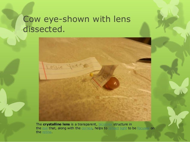
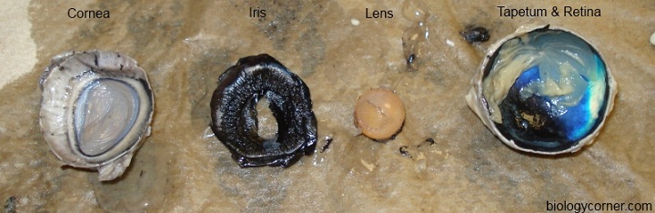
Describe the Lens of the Dissected Cow Eye
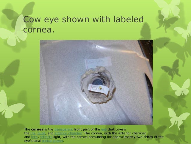
Cornea - tough covering over the iris that helps protect the eye and directs the light towards the lens. Learn how to dissect a cows eye in your classroom.
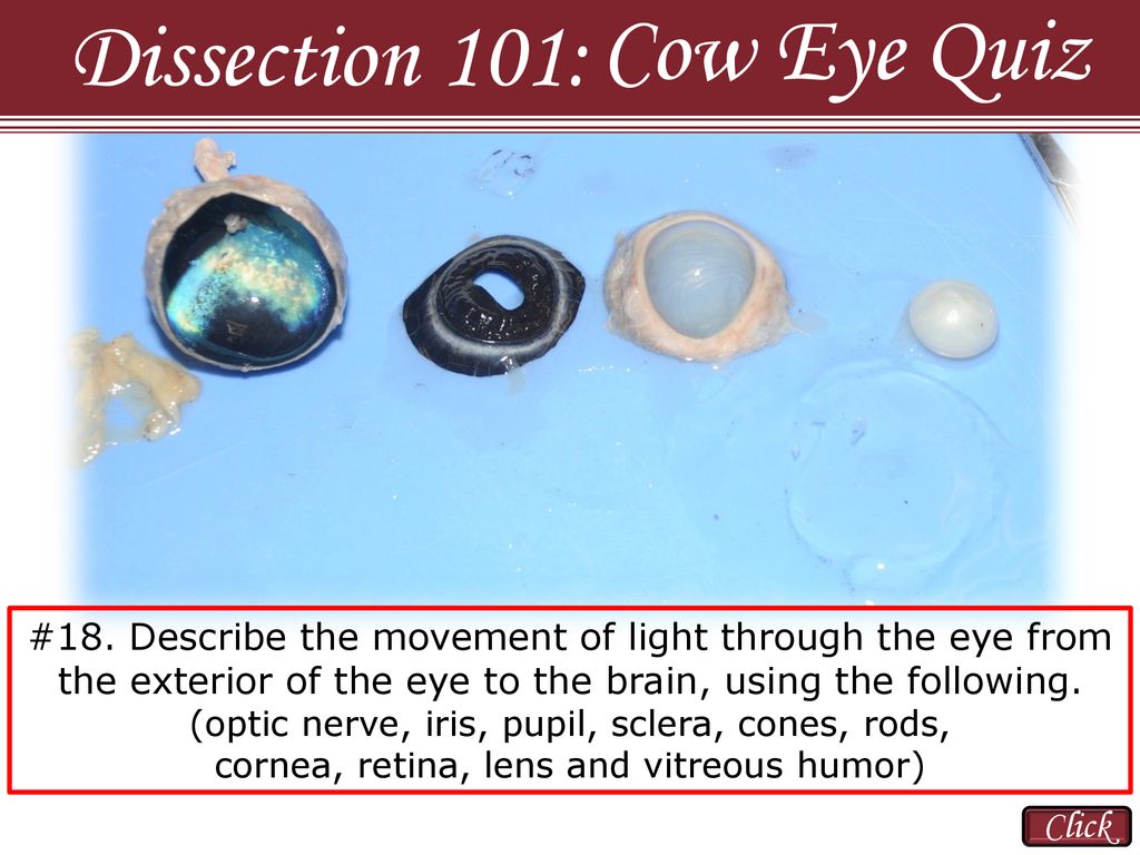
Cow Eye Quiz Dissection 101 Click Ppt Download
Use the structures listed in question 2 and label the diagram.

. Sends messages from the eye to the brain. During this activity you will dissect a cow eye. Outer lens that lets light into the eye.
Up to 24 cash back 2. Describe the image of the student as you look through. The eye is the organ of sight.
A ring of muscle tissue that forms the colored portion of the eye. Describe the vitreous humor of the dissected eye. The parts we will be examining are the cornea sclera optic nerve iris lens pupil ciliary body and aqueous humor.
The human pupil is rounded. Up to 24 cash back The four muscles that moves the cows eye in four directions. Colored ring of muscle that changes the size of the pupil.
O The lens of the cows eye feels soft on the outside and hard in the middle. Examine the outside of the eye. Optic nerve- The bundle of.
The transparent structure behind the pupil that changes shape to help focus images on the retina. This tough outer covering of the eyeball has fat and muscle attached to it. A covering over the iris that helps to protect the eye by making light bend to project the image.
As you get closer to the actual eyeball you may notice muscles that are attached directly to the sclera and along the optic nerve. There are many parts of the eye that all play a part in helping us see. Materials Preserved Cow Eye Scalpel or Scissors Forceps Dissection Tray Gloves Safety Glasses Lab Apron 1.
Describe the tissue of the lens you felt during the dissection of the cow eye. This portion of the lab is based on one used in the Introductory Physics course. What is the function of the dark pigment in the choroid coat.
Optic nerve iris pupil sclera cones rods cornea retina lens and vitreous humor Use a labeled drawing if it is. When the cow was alive the cornea was clear. What is the function of the lens.
It felt hard like a marble but it was not shaped exactly spherical like a marble. O The lens is a transparent sphere that can change its shape to focus the light entering the eye. Place the cows eye on a dissecting tray.
While the cow was alive the lens was clear and very flexible. Get the cow eye. Describe the lens of the dissected eye.
Lab Virtual Eye Dissection. You should be able to find the sclera or the whites of the eye. The white part of the eye the sclera is a tough outer covering of the eyeball.
Gloves Cow eye Warm water Paper towel Dissection pan Scalpel Scissors Probe Procedure. The Cows Eye httpswwwaxploratorium edullideolcow-eye-dissection Step 1 and 2 1. You will need a scalpel and forceps.
Controls the size of the pupil opening. DISSECTION OF THE COW EYE Please make sure to wear gloves and safety glasses when you are dissecting and make sure to clean up thoroughly after the lab. Insert sciss the way are it into ante As you ide anterior p then write Locate on a dissected cow eye and describe the function of each of the following structures.
After examining both sides of the anterior half of the eye remove the lens out by cutting the ciliary body with your scissors. Together with the lens the cornea refracts light and helps the eye to focus. Below is an example of a response for light movement through the eye.
A viscous liquid that provides shape to the eye. Extrinsic eye muscle optic nerve sclera cornea pupil lens iris ciliary body choroid retina optic disc vitreous humor and tapetum lucidum Materials Needed Fresh or preserved cow eye Lens Dissection tray Dissection. The tissue in the lens of the cow eye was hard and tough.
Why is the vitreous humor clear. Describe the movement of light through the eye from the exterior of the eye to the brain using the following. These are the extrinsic muscles that allow a cow to move its eye up and down and from side to side.
Locate the covering over the front of the eye the. The purpose of this section of the lab is to use the lens dissected from the fresh cow eye to investigate the physics of the lens specifically the relationship between image and object distance as they relate to focal length for different lenses. Also the cow eyes can be rather slippery so use caution when handling and cutting them.
As we have seen in the dissection a cows iris is. The blue covering over the front of the eye is the cornea. Carefully cut away the fat and the muscle.
Up to 24 cash back a. A viscous liquid that provides shape to the eye. It felt hard like a marble but it was not shaped exactly spherical like a marble.
Located in the back of the eye contains the rods and cones. Biconvex flexible and transparent. Location of the retina where there are no rods or cones.
First identify the most external structures of the eye. Optic nerve iris pupil sclera cones rods cornea retina lens and vitreous humor Use a labeled drawing if it is helpful. Describe the function of the following structures.
Ask the students what they see when they hold up the lens and look through it They see. Absorbs excess light and keeps the eye dark inside. Depending on the breadth of students cow eye samples students can identify both internal and external parts of the eye.
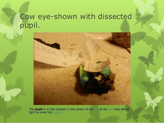
Hole in the iris that allows light into the inner eye. Rinse with the water in your tray and pat dry with a paper towel. The pupil of the dissected eye was an elliptical shape.
The iris is a muscle that controls how much light goes into the eye and suspended between the cornea and lens. The tissue in the lens of the cow eye was hard and tough. Describe the movement of light through the eye from the exterior of the eye to the brain using the following.
Step 7 12 What to the poly tko substance that sunround the tena. Anatomy of a Cows Eye. The eye most likely has a thick covering of fat and muscle tissue.
In a preserved cow eye the lens will most likely have yellowed and become very hard. You will observe several important features of the eye and develop your understanding of how each part functions to make vision possible. A step-by-step hints and tips a cow eye primer and a glossary of terms.
Describe the tissue of the lens you felt during the dissection of the cow eye. Identify the following structures and describe their function. The sclera gives the eye its shape and helps to protect the delicate inner parts.
By examining a cows eye students learn the parts of the eye and the structures that surround it. Students can also recognize distinct structures like the pupil cornea optic nerve lens and iris.
Biology Lab 10 Cow Eye Dissection Diagram Quizlet

Cow Eye Dissection Cows Compared To Humans Without Moving Your Head Look Up Look Down Look All Around Six Muscles Attached To Your Eyeball Move Ppt Download
Cow Eye Dissection And Label
Cow Eye Dissection And Label
Cow Eye Dissection And Label

Cow Eye Dissection Perkins Elearning
Cow Eye Dissection And Label

Carolina S Perfect Solution Preserved Cow Eyes Carolina Com

580181 Mammalian Eye Dissection Ada Pdf Anatomy Physiology Mammalian Eye Dissection Investigation Manual Mammalian Eye Dissection Table Of Contents 2 Course Hero

Frog Anatomy Review Pig Dissection Dissection Animal Science
Eye Dissection Ppt Download

Eye Dissection The Eyes Have It Edu Youtube

Eye Dissection Name Period Ppt Download

Cow S Eye Dissection Exploratorium Video
Cow Eye Dissection Carolina Com
Cow Eye Dissection And Label

Cow Eye Dissection Parts Of The Eye Diagram Quizlet
Cow Eye Dissection
The Eye An Amazing Optical System


Comments
Post a Comment